
Un método rápido y efectivo para preparar hormigas para microscopía electrónica de barrido
Barra lateral del artículo
Contenido principal del artículo
Resumen
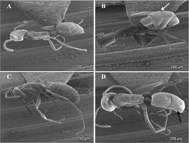
Independientemente de la aplicación de técnicas de microscopía electrónica de barrido (MEB), la preparación adecuada del material biológico a analizar es de suma importancia. En la mayoría de los estudios de hormigas que involucran SEM, las hormigas han sido desecadas por la técnica de punto crítico (CPD por sus siglas en inglés), pero la mayoría de las especies de Dolichoderinae tienen tegumentos delgados y, por lo tanto, tienden a colapsar fácilmente. Para evaluar las ventajas potenciales de un nuevo método sobre la técnica CPD, estas hormigas de tegumentos delgados se trataron con tetrametilsilano (TMS) y luego se secaron al aire. Los resultados obtenidos en este estudio se presentan en microfotografías de barrido. Aquí, detallo un protocolo estandarizado para la preparación de hormigas con TMS antes de SEM. La técnica TMS permite el análisis de casi cinco a seis veces más hormigas que CPD y es más rápido, más fácil, más eficiente y económico que el método CPD.
Descargas
Detalles del artículo

Esta obra está bajo una licencia internacional Creative Commons Atribución-NoComercial-CompartirIgual 4.0.
Citas
Barré, C., O’neil D. and Bricelj, V.M. 2006. Preparation of large bivalve specimens for scanning electron microscopy using Hexamethyldisilazane (HMDS). Journal of Shellfish Research 25: 639 - 641. Doi: https://doi.org/10.2983/0730-8000(2006)25[639:POLBSF]2.0.CO;2.
Baroni Urbani, C. and De Andrade, M.L. 2007. The ant tribe Dacetini: Limits and constituent genera, with descriptions of new species. Annali del Museo Civico di Storia Naturale Giacomo Doria (Genova) 99: 1-191.
Botes L, Price, B., Waldron, M. and Pitcher, G.C. 2002. A simple and rapid scanning electron microscope preparative technique for delicate ''Gymnodinioid'' dinoflagellates. Microscopy Research and Technique 59: 128 - 130. Doi: https://doi.org/10.1002/jemt.10184.
Bozzola, J.J. and Russel, L.D. 1998. Electron Microscopy. Principles and Techniques for Biologists, Second edition. Jones and Bartlett publishers, Sudbury, Massachusetts.
Bray, D.F., Bagu J. and Koegler, P. 1993. Comparison of hexamethyldisilazane (HMDS), Peldri II, and critical point drying methods for scanning electron microscopy of biological specimens. Microscopy Research and Technique 26:489-495. Doi: https://doi.org/10.1002/jemt.1070260603.
Camargo-Mathias, M.I., Fantazzini, E.R., Fontanetti, C.S. and Calligaris, I.B. 2011. 3D reconstruction and scanning electron microscopy of salivary glands of the millipede Rhinocricus padbergi (Verhoef, 1938) (Diplopoda: Spirobolida). Micron 42: 271-274. Doi: https://doi.org/10.1016/j.micron.2010.10.004.
Cuezzo, F. and Guerrero, R.J. 2012. The ant genus Dorymyrmex Mayr (Hymenoptera: Formicidae: Dolichoderinae) in Colombia. Psyche 2012: 1-24. Doi: https://doi.org/10.1155/2012/516058.
Dahl, C. 1972. Preparation of alcohol preserved larvae of culicidae for scanning electron microscopy. Entomological Scandinavia 3: 181-188. Doi: https://doi.org/10.1163/187631272X00283.
De Andrade, M. and Baroni Urbani, C. 1999. Diversity and adaptation in the ant genus Cephalotes, Past and Present. Stuttgarter Beiträge zur Naturkunde Serie B (Geologie und Paläontologie) 271: 1-889.
Dey, S. 1993. A new rapid method of airdrying for scanning electron microscopy using tetramethylsilane. Application to mammalian tissue. Cytobios 73: 17-23. Doi: https://doi.org/10.1111/j.1365-2818.1989.tb02925.x.
Dey, S., Basu-Baul, T.S., Roy B. and Dey, D. 1989. A new rapid method of airdrying for scanning electron microscopy using tetramethylsilane. Journal of Microscopy 156: 259-261. Doi: https://doi.org/10.1111/j.1365-2818.1989.tb02925.x.
Fernández, F. 2004. Adelomyrmecini new tribe and Cryptomyrmex new genus of myrmicine ants (Hymenoptera: Formicidae). Sociobiology 44: 325-335.
Fox E.G.P., Bueno, O.C., Yabuki, A.T., Jesus, C.M., Solis, D.R., Rossi M.L. and Nogueira, N.L. 2010. General morphology and ultrastructure of the venom apparatus and convoluted gland of the fire ant, Solenopsis saevissima. Journal of Insect Science 10: 24. Doi: https://doi.org/10.1673/031.010.240.
Guerrero, R.J. and Fernandez, F. 2008. A new species of the ant genus Forelius (Formicidae: Dolichoderinae) from the dry forest of Colombia. Zootaxa 1958: 51-60. Doi: https://doi.org/10.11646/zootaxa.1958.1.5.
Gorb, E., Haas, K., Henrich, A., Enders, S., Barbakadze, N. and Gorb, S. 2005. Composite structure of the crystalline epicuticular wax layer of the slippery zone in the pitchers of the carnivorous plant Nepenthes alata and its effect on insect attachment. The Journal of Experimental Biology 208: 4651-4662. Doi: https://doi.org/10.1242/jeb.01939.
Hita Garcia, F., Fischer, G., Liu, C., Audisio, T.L. and Economo, E.P. 2017. Next-generation morphological character discovery and evaluation: An X-ray micro-CT enhanced revision of the ant genus Zasphinctus Wheeler (Hymenoptera, Formicidae, Dorylinae) in the Afrotropics. ZooKeys 693: 33-93. Doi: https://doi.org/10.3897/zookeys.693.13012.
Jung, S.W., Joo, H.M., Park, J.S. and Lee, J.H. 2010. Development of a rapid and effective method for preparing delicate dinoflagellates for scanning electron microscopy. Journal of applied phycology 22: 313-317. Doi: https://doi.org/10.1007/s10811-009-9461-6.
Keller, R. 2011. A phylogenetic analysis of ant morphology (Hymenoptera: Formicidae) with special reference to the Poneromorph subfamilies. Bulletin of the American Museum of Natural History 355: 1-90. Doi: https://doi.org/10.1206/355.1.
Laforsch, C. and Tollrian, R. 2000. A new preparation technique of daphnids for Scanning Electron Microscopy using hexamethyldisilazane. Archiv für Hydrobiologie 149: 587-596. Doi: https://doi.org/10.1127/archiv-hydrobiol/149/2000/587.
Lattke, J.E. 2000. Specimen processing: Building and curating an ant collection. In: Agosti, D. Majer, J.D., Alonso L.E. and Schultz T.R., Editors. Ants, Standar Methods for Measuring and Monitoring Biodiversity. Smithsonian Institution Press, Washington, D.C.
Lee, P. and Szema, R. 2005. Inspirations from biological optics for advanced photonic systems. Science 310: 1148-1150.
Doi: https://doi.org/10.1126/science.1115248.
Lucas, C., Fresnau, D., Kolmer, K., Heinze, J., Delabie, J.C.H. and Pho, D.B. 2002. A multidisciplinary approach to discriminating different taxa in the species complex Pachycondyla villosa (Formicidae). Biological Journal of the Linnean Society 75: 249-259. Doi: https://doi.org/10.1111/j.1095-8312.2002.tb01425.x.
Mehdizadeh, K.A., Tahermanesh, K., Chaichian, S., Joghataei, M.T., Moradi, F., Tavangar, S.M., Najafabadi, A.S.M., Lotfibakhshaiesh, N., Beyranvand, S.P., Anvari-Yazdi, A.F and Abed, S.M. 2014. How to prepare biological samples and live tissues for scanning electron microscopy (SEM). Galen Medical Journal 3(2): 63-80. Doi: https://doi.org/10.31661/gmj.v3i2.267.
Nation, J.L. 1983. A new method using hexamethyldisilazane for preparation of soft insect tissues for scanning electron microscopy. Stain Technology 58: 347-351. Doi: https://doi.org/10.3109/10520298309066811.
Reville, W.J. and Cotter, M.P. 1991. An evaluation of the usefulness of air-drying biological samples from tetramethylsilane in preparation for scanning electron microscopy. Journal of Electron Microscopy 40: 198-202. Doi: https://doi.org/10.1093/oxfordjournals.jmicro.a050896.
Ting-Beall, H.P., Zhelev, D.V. and Hochmuth, R.M. 1995. Comparison of different drying procedures for scanning electron microscopy using human leukocytes. Microscopy Research and Technique 32: 357-361. Doi: https://doi.org/10.1002/jemt.1070320409.
Ubero-Pascal, N.J. and Puig, M.A. 2007. Egg morphology update based on new chorionic data of Potamanthus luteus (Linnaeus), Ephemera danica Müller and Oligoneuriella rhenana (Imhoff) (Insecta, Ephemeroptera) obtained by scanning electron microscopy. Zootaxa 1465: 15-29. Doi: https://doi.org/10.11646/zootaxa.1465.1.2.
Ubero-Pascal, N.J. and Puig, M.A. 2009. New type of egg attachment structure in Ephemeroptera and comparative analysis of chorion structure morphology in three species of Ephemerellidae. Acta Zoologica 90: 87-98. Doi: https://doi.org/10.1111/j.1463-6395.2008.00367.x.
Ubero-Pascal, N.J., Fortuño, M. and Puig, M.A. 2005. New application of air-drying techniques for studying Ephemeroptera and Plecoptera eggs by scanning electron microscopy. Microscopy Research and Technique 68: 264-271. Doi: https://doi.org/10.1002/jemt.20248.
Vukusic, P., Sambles, J.R., Lawrence, C.R. and Wootton, R.J. 1999. Quantified interference and diffraction in single Morpho butterfly scales. Proceedings of the Royal Society London Series B 266: 1403-1411. Doi: https://doi.org/10.1098/rspb.1999.0794.
Yaws, C.L. 2014. Thermophysical properties of chemicals and hydrocarbons. Second edition. Elsevier press, Oxford.